

SPOTLIGHT
HOW SMART-C® SUPPORTED COMPLEX VETERINARY TRAUMA SURGERY
Dakota, a five-year-old Boxer struck by a vehicle, underwent advanced diagnostic imaging and orthopedic repair supported by SMART-C® real-time fluoroscopic guidance.
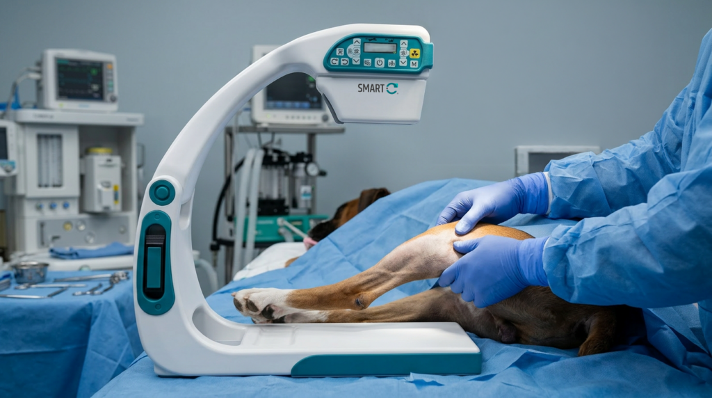
Patient: Dakota, five-year-old Boxer
Setting: Emergency veterinary hospital
Condition: Open left tibial fracture following vehicle trauma
Imaging need: Real-time surgical guidance
Challenge: Accurate fracture reduction and alignment during complex orthopedic repair
Dakota was brought in for emergency evaluation after being struck by a moving vehicle. Diagnostic imaging revealed an open compound fracture of the left tibia, along with additional injuries associated with blunt force trauma.
Following stabilization and comprehensive imaging, the veterinary team used SMART-C to support surgical planning, fracture assessment, and real-time intraoperative guidance during orthopedic repair.
THE CHALLENGE
Complex veterinary trauma cases require rapid diagnostics, careful surgical planning, and immediate imaging support.
Dakota’s injuries required accurate fracture reduction, alignment assessment, and intraoperative visualization while minimizing surgical risk and the potential need for revision procedures.
SMART-C® provided real-time fluoroscopic imaging directly within the procedural environment, helping the surgical team evaluate positioning dynamically throughout the repair.
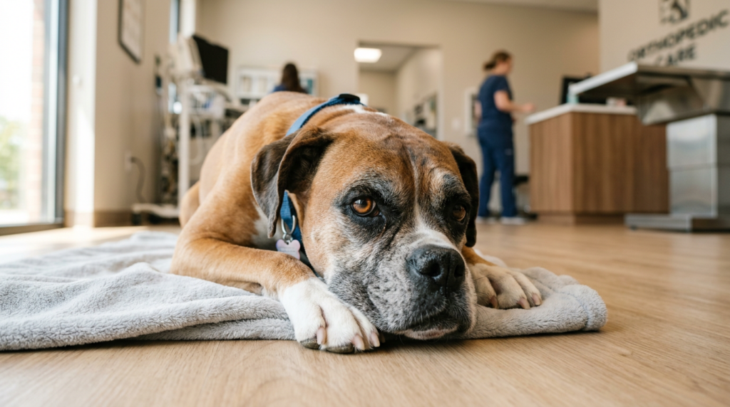
“SMART-C enabled real-time assessment of alignment and joint mobility during fracture repair, supporting procedural confidence and eliminating the need for revision.”
SMART-C® CLINICAL AND OPERATIONAL IMPACT
During orthopedic intervention, SMART-C® provided real-time visualization throughout fracture reduction and repair. Fluoroscopic imaging supported anatomical alignment, procedural accuracy, and intraoperative decision-making.
Continuous visualization allowed the veterinary team to confirm alignment, assess joint positioning, and evaluate mobility without interrupting surgical workflow or repeatedly repositioning the patient.
At the veterinary hospital, SMART-C® supported:
- Surgical planning and hardware placement
- Real-time fracture reduction
- Intraoperative alignment assessment
- Dynamic joint and mobility evaluation
- Imaging without repeated patient repositioning
Following surgery, Dakota recovered successfully with continued monitoring, pain management, and supportive care.
REAL-TIME
Fluoroscopic imaging guidance
DYNAMIC
Intraoperative alignment assessment
PRECISE
Fracture reduction visualization
CONTINUITY
No repositioning during imaging
Get in Touch
We’ll follow up shortly to learn more about your needs.
Capabilities and workflows may vary based on state regulations and facility protocols.
Follow federal, local, and institutional regulations for X-ray imaging use and radiation safety protection.